Bunion
Correction.
Relieve pain and restore your foot's natural alignment with Peoria's leader in advanced bunion surgery and minimally invasive techniques.
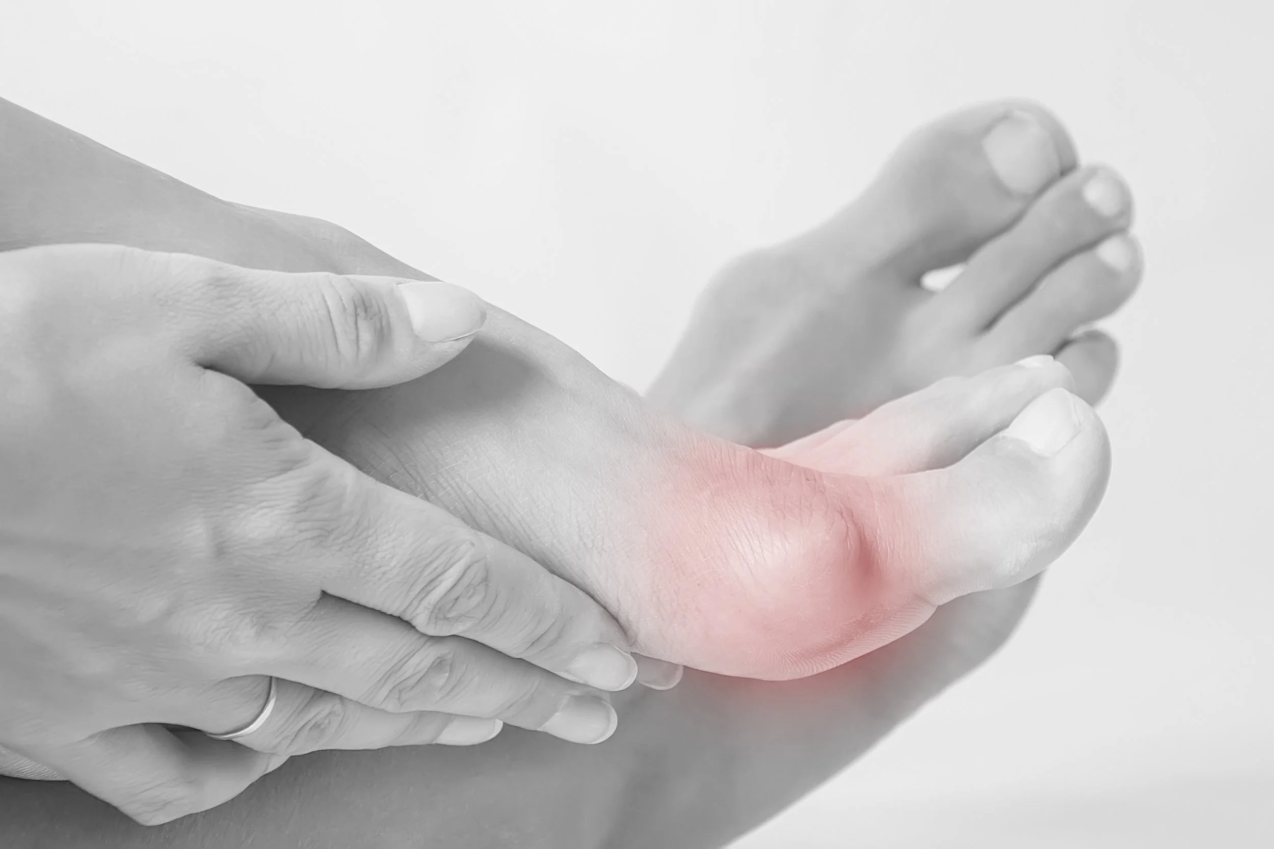
What is a Bunion?
A bony bump that forms on the joint at the base of your big toe, occurring when some of the bones in the front part of your foot move out of place.
Common Symptoms
Pain, swelling, redness, and a visible bump. Often leads to stiffness and difficulty wearing regular shoes.
The Expert Approach
Dr. Kaveh Panahi utilizes 4th generation minimally invasive surgical techniques to prioritize faster recovery and aesthetic outcomes.
Minimally Invasive
Bunionectomy (MICA)
Dr. Panahi specializes in the 4th Generation MICA (Minimally Invasive Chevron/Akin) procedure. This advanced technique corrects the deformity through tiny openings rather than large incisions.
Faster Recovery: Get back on your feet significantly sooner than traditional "open" surgery.
Minimal Scarring: Tiny incisions mean superior aesthetic results.
Less Pain: Reduced trauma to soft tissues and blood vessels during the correction.
The MICA Procedure Step-by-Step
Bunion
A bunion is a bony bump at the base of the big toe that forms when the toe shifts inward. This misalignment causes pain, swelling, and discomfort, especially in shoes, making walking and daily activities difficult. Treating bunions early can relieve pain and prevent worsening issues.

Incision
MIS bunion surgery uses tiny incisions, typically less than 0.5 cm, to access and correct the bunion. These small cuts minimize damage to surrounding tissues, reduce scarring, and lead to faster healing with less post-operative pain. This approach allows for a quicker recovery and a more comfortable return to daily activities.
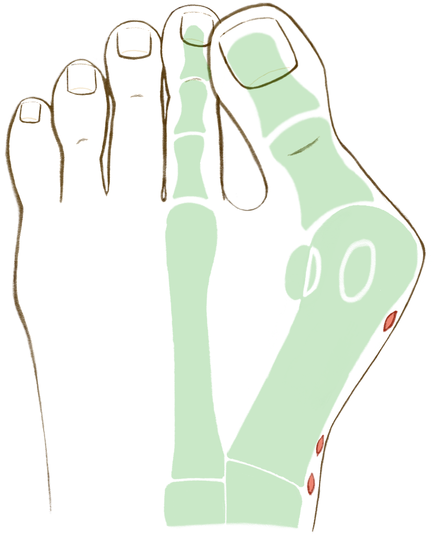
Correction
The head of the metatarsal bone is precisely shifted into a corrected position to realign the big toe. Two small but strong titanium screws are used to secure the bone in place, providing stability while allowing for weight bearing. These screws ensure long-lasting correction while promoting quicker healing and improved foot function.
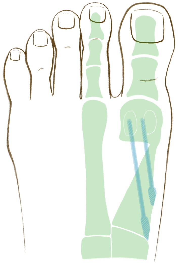
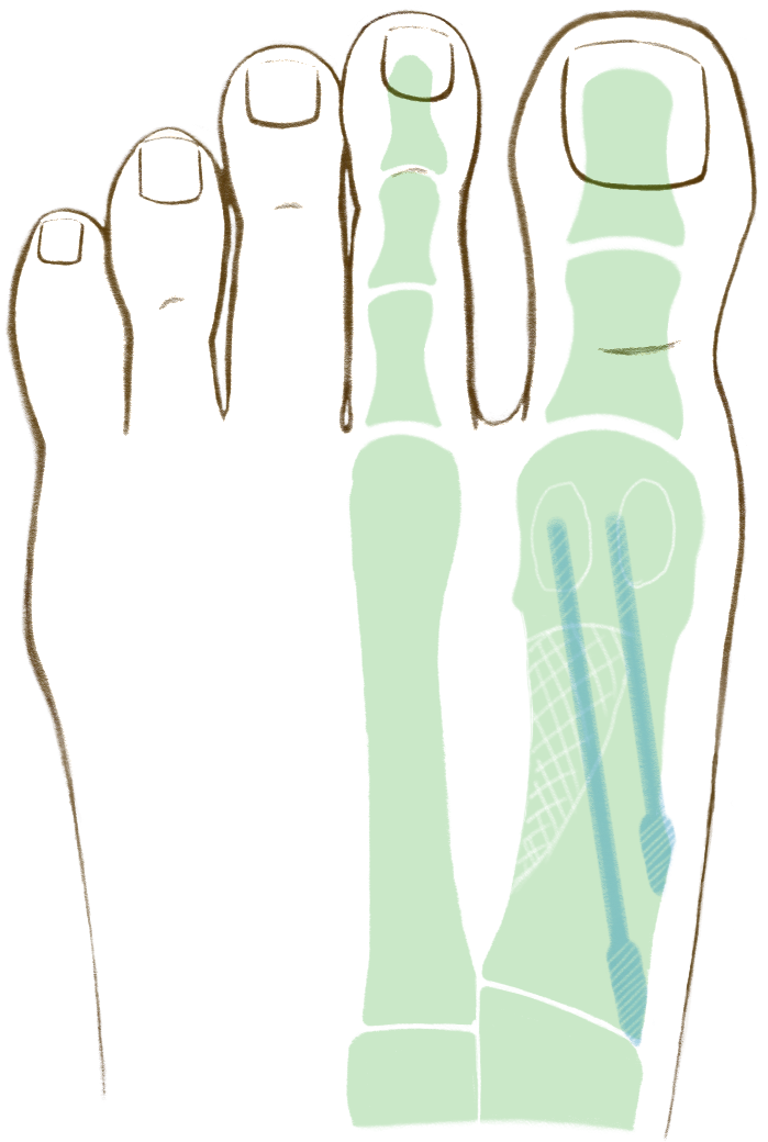
Healing
The gap created by shifting the bone gradually fills with new bone over the next several weeks, promoting a stable, natural healing process. Most patients can start walking in a protective boot within the first few days, allowing for gentle mobility while the bone heals. This early walking aids recovery, helping patients return to normal activities sooner with minimal discomfort.
Video Animation of MIS Bunion Correction
Precision Surgical Techniques
Every foot is unique. We offer a full spectrum of surgical corrections.
Lapidus (Lapiplasty 3D Bunionectomy)
The Lapiplasty procedure corrects bunions by fusing the joint between your first metatarsal bone and one of the small bones in the middle of your foot — the medial cuneiform. Screws or plates fuse your bones together — this helps to straighten your big toe and prevent future bunions. The procedure can also correct your bunion in three dimensions, fixing any angles or rotations that cause you problems or pain.
Austin (Chevron) Bunionectomy
An Austin Bunionectomy is a surgical procedure used to correct mild to moderate bunions by realigning the big toe joint. This technique involves making a V-shaped cut (known as a distal metatarsal osteotomy) in the first metatarsal bone to shift it into a more natural position. The bone is then stabilized using a small surgical screw or pin. The goal of the procedure is to reduce pain, improve foot function, and restore a more natural foot shape—all while preserving joint movement. Recovery typically involves a protective boot and gradual return to normal activity under your surgeon’s guidance.
Non-Surgical Path
We typically encourage conservative, non-surgical treatments before considering surgery. Dr. Panahi will conduct a physical exam and X-ray to determine the severity of your condition.
- Custom Orthotics & Inserts
- Bunion Pads & Toe Spacers
- Anti-inflammatory Management
- Specialized Stretches & Exercises
Your Consultation
During your initial visit, Dr. Panahi uses high-resolution digital imaging to map the structural alignment of your foot. We don't just look at the bump; we analyze the mechanical cause to ensure your treatment plan provides lasting relief and prevents recurrence.
Analysis
Weight-bearing X-rays to assess the degree of deformity.
Custom Plan
Tailored recovery timeline based on your lifestyle and activity level.
*Results and recovery times may vary depending on the individual patient and specific surgical technique utilized.